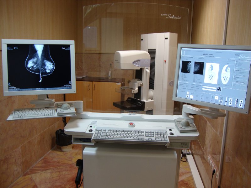
With current advantages in digital
imaging systems especially digital mammography great change in the diagnosis and
follow up of breast cancer has been occurred. In the Haghighat Imaging Center
breast imaging department the most sophisticated and modern Digital mammography
from the Hologic company has been implemented which is FDA approved . Not only
the digital mammography increases the diagnostic accuracy but also the patient
has less pain and less X-ray exposure. All the images are reported in the
standard monitors with ability to transfer to the PACS system and save the
images for future comparative studies. They are reported by the well skilled
team who are expert in breast imaging. It is also possible to perform screening
mammography with this system and save for the future comparison. Preparation for
mammography;
- Second week of menstruation is preferred because pain is least and the
cell nuclei are less susceptible to X-ray. Also possibility of pregnancy is
almost excluded in this time.
- Remember to bring the previous documents. Especially previous
mammographic images.
- Take a shower in the same day and avoid using deodorant.
Breast Ultrasonography
The breast ultrasonography studies is done by Mylab 60 and from Esoate company
With dedicated probe for breast imaging. It has high resolution with ability to
detect superficial as well as deep retro mammary spaces and excellent quality.
Vascular status can be evaluated by color Doppler. Also the breast Biopsy
including core needle biopsy and vacuum assisted device is available. Wire
localization of lesions with sonographic guide is done as well.
Biopsy preparation
- After the first presentation, the patient's documents will be evaluated
by radiologist. A control sonography might be necessary before date of biopsy.
- Anticoagulant therapy should be stopped for 1-2 days
- Anesthesia is done in two stages, subcutaneous with very thin needle and
deeper with thicker needle.
- According to the type of lesion and clinicians order, biopsy might be
performed with core needle device or vacuum assisted machine.
- The samples might be checked by X-ray if necessary.
- Samples in formalin will be referred to pathologist.
MR Mammography with and without contrast
It is a safe method of evaluation of breast with MRI .In this method a dedicated
coil for breast is used and by skilled team related sequences acquired. All the
images transfer to the PACS system and evaluated in specialized work station.
The dynamic curves are obtained by the radiologist herself. Comparison with
patient's history and documents including previous mammography and
ultrasonography is done as well.
Breast MRI Preparation:
- This is necessary to perform Breast imaging especially MRI in the second
week of menstrual period to avoid hormonal unpleasant interaction.
- The patient lies prone and her breasts are hanged in coils without
pressure. Breathing is easy possible because the forehead is leaning and face is
located in a free space.
- IV line is available
- The patient might use headphone to be more comfortable.
- Precontrast images including 2-3 series will be obtained.
- The technician will come in the room and injection will be performed.
- 6 series of same images will be obtained consequently.
- The emergency cases will be evaluated in the same day.
- The prostheses are evaluated with specific sequences.